

https://doi.org/10.3389/fpls.2022.1036078
“Sap flow measurement is one of the most effective methods for quantifying plant water use.A better understanding of sap flow dynamics can aid in more efficient water and crop management, particularly under unpredictable rainfall patterns and water scarcity resulting from climate change. In addition to detecting infected plants, sap flow measurement helps select plant species that could better cope with hotter and drier conditions. There exist multiple methods to measure sap flow including heat balance, dyes and radiolabeled tracers. Heat sensor-based techniques are the most popular and commercially available to study plant hydraulics, even though most of them are invasive and associated with multiple kinds of errors. Heat-based methods are prone to errors due to misalignment of probes and wounding, despite all the advances in this technology. Among existing methods for measuring sap flow, nuclear magnetic resonance (NMR) is an appropriate non-invasive approach. However, there are challenges associated with applications of NMR to measure sap flow in trees or field crops, such as producing homogeneous magnetic field, bulkiness and poor portable nature of the instruments, and operational complexity. Nonetheless, various advances have been recently made that allow the manufacture of portable NMR tools for measuring sap flow in plants. The basic concept of the portal NMR tool is based on an external magnetic field to measure the sap flow and hence advances in magnet types and magnet arrangements (e.g., C-type, U-type, and Halbach magnets) are critical components of NMR-based sap flow measuring tools. Developing a non-invasive, portable and inexpensive NMR tool that can be easily used under field conditions would significantly improve our ability to monitor vegetation responses to environmental change.”
”
Nuclear magnetic resonance for sap flow measurements
Nuclear magnetic resonance (NMR) is a phenomenon when a strong magnetic field and the nucleus-based phenomenon is basically related to the absorption and re-emission of electromagnetic wave radiation. This phenomenon was introduced in 1938 (Rabi et al., 1938; Rabi et al., 1939). In the early 1940s, Felix Bloch and Edward Purcell independently developed the NMR method for measuring nuclear magnetic moments, for which they shared the Nobel Prize in 1952. NMR applications developed rapidly over the next 50 years. Nuclei with odd numbers of protons and neutrons have non-zero spins, making them act as tiny bar magnets. Their orientation is normally random and there is no net magnetic field, so they cannot generate an NMR signal. In NMR, when these nuclei are subjected to an external strong and homogenous magnetic field, nuclei spins are orientated along the magnetic field direction; some in parallel (low energy) and some others in antiparallel directions (high energy). This results in a net magnetic moment generated which is aligned with the external magnetic field. This net magnetization can be pushed away from the alignment using a second magnetic field applied perpendicular to the external magnetic field with an excitation coil. Net magnetization returns to the external magnetic field when this second field is turned off, generating a radiofrequency signal which is detected at the detector coil. This signal forms the basis of NMR imaging and spectroscopy.
The energy difference between the two states of nuclei spin under an external strong magnetic field is very small and corresponds to a range of frequency of the EM spectrum. A short duration radio frequency signal is used to flip the nuclear spin from the spin-aligned state of low energy to the spin-opposed state of high energy. Thereafter, the charged nucleus will exhibit processional motion with a specific frequency, known as the Larmor frequency. The actual magnetic field created at a nucleus is also dependent on the electrons surrounding it, which creates an ionic environment around it which leads to a variation in the frequency of resonating particles, known as chemical shift. This change magnetic field is known as shielding. Energy is absorbed by the nuclei during the transition from lower to higher energy levels and released during the opposite process. The process of returning to the original energy state is known as relaxation. The two most common types of relaxation are spin lattice relaxation (T1) and spin relaxation (T2). T1 relaxation, also known as spin lattice or longitudinal relaxation is the time constant used to describe when ~63% of the magnetization has recovered to equilibrium. T2 relaxation involves energy transfer between interacting spins via dipole and exchange interactions. Spin-spin relaxation energy is transferred to a neighboring nucleus. Now there are two NMR methods continuous and pulsed method. In continuous wave NMR method, a sample in a static magnetic field is applied by RF perpendicular to the sample and when an excitation frequency is reached, oscillation is observed at the detector. In the pulsed case, one applies a sequence of RF pulses called π-pulses (180-degree pulses) or π/2-pulses (90-degree pulses) and looks for “free induction decay” and “spin echoes”. Use of Pulse NMR experiments are most popular these days.
Capturing all details of these reaction can be conducted by accurate measurement systems. The captured waveforms which are known as NMR signals, release fundamental features regarding the understudy materials; see Figure 3 (Callaghan et al., 1991). Concept of NMR has been applied in many research areas e.g., in plant studies to determine moisture in apple (Shaw and Elsken, 1956), unfrozen water in winter cereals at subfreezing temperatures (Gusta et al., 1975), and xylem embolisms in pine wilt disease (Umebayashi et al., 2011). One advantage of NMR is that it can spatially resolve both static and dynamic parameters and generate data in a non-destructive manner from the interior of a sample.
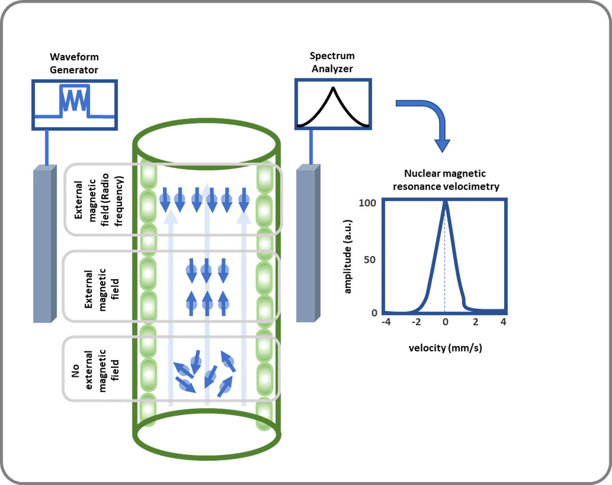 FIGURE 3 This diagram illustrates how SAP flow is measured using NMR. SAP flow measurement using NMR includes a strong magnetic field where 1H nucleus gains a quantum mechanical property callspin at. Then an NMR signal is created by applying RF energy equal to the Larmor frequency, which leads to nuclei transitioning from their groan und to excited state, and spins being oriented oppositely or antiparallelly. After receiving, amplifying, and further analyzing the signal, the volumetric flow rate of the xylem and phloem is calculated.
NMR can be used in different applications where non-destructive measurements are required. NMR imaging is also widely used to measure sap flow (Table 2). van As and Schaafsma (1984) investigated water flow through the stem of an intact cucumber plant using pulsed NMR. In their study, the NMR signal due to high stationary water was cancelled out so that it doesn’t interfere in the detection of flowing water. This made it an important application for measuring water flow to plants with high stationary water content. Using NMR microimaging, Köckenberger et al. (1997) measured the phloem and xylem water flow in castor bean seedlings. This study demonstrated for the first time that water is internally circulated between phloem and xylem. Even in absence of significant transpiration or evaporation, there is water maintenance through this water circulation. Fast gradient echo sequence (FLASH) imaging of flow velocities, as a fast-imaging NMR method, was introduced by Rokitta et al. (1999). Their method achieved a six-fold reduction in imaging time while maintaining a similar signal-to-noise ratio to previous flow NMR imaging techniques. Scheenen et al. (2000) developed the pulsed field gradient (PFG) NMR technique combined with turbo spin-echo imaging, which resulted in an accurate and spatially resolved sap flow measurement. Windt et al. (2006) used NMR imaging for diurnal dynamics study of phloem and xylem transport in poplar (Populus spp), castor bean (Ricinus communis), tomato (Solanum lycopersicum) and tobacco (Nicotiana tabacum). They found large diurnal variation in xylem flux and small or no change in phloem flux.
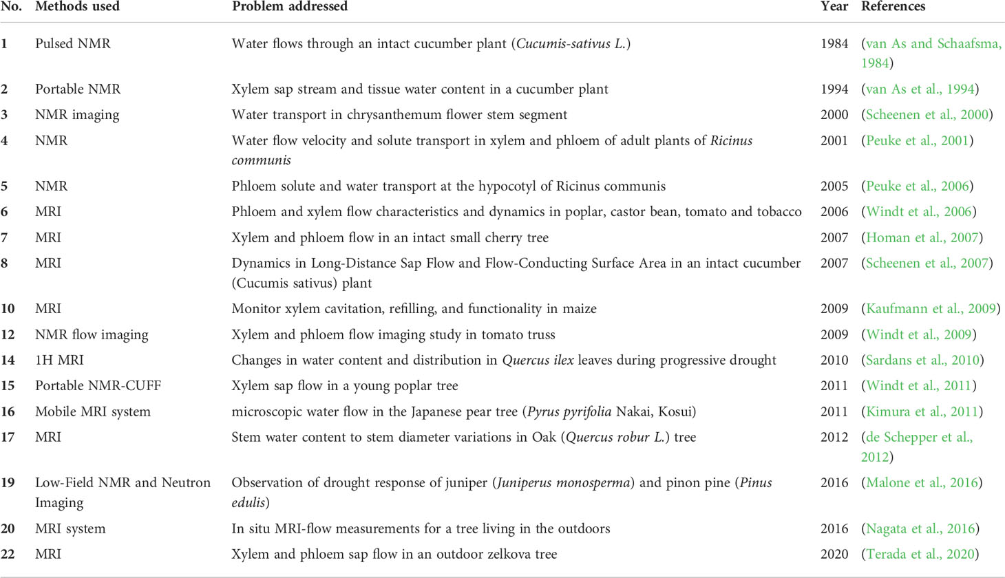 TABLE 2 Different studies used NMR and other imaging methods to study sap flow measurement in plants and trees.
Using NMR flow imaging, Windt et al. (2009) demonstrated that, in a developing tomato truss, 75% of net influx into a fruit occurred through the xylem and the remaining 25% via the perimedullary region, which includes both phloem and xylem. Using NMR methodology, the moisture content in wood samples could be instantly determined from the mass and amplitude of free-induction-decay (FID) signals and found this methodology to be more precise and reliable than the gravimetric methods regardless of wood species (Merela et al., 2009). Furthermore, NMR imaging can determine the level of hydraulic conductivity in detached stems. Using MRI, de Schepper et al. (2012) showed that elastic bark tissues contribute most to the daily depletion of internal stem water storage. In another study, low field NMR and neutron imaging were used to deduce aspen tree (Populus tremuloides) drought response (Malone et al., 2016). NMR imaging was also used for grape (Vitis vinifera) stems to show that basal leaves were more prone to embolism than apical leaves and these leaves shed to prevent water loss and protect the hydraulic integrity of the plant (Hochberg et al., 2017).
The variety of possible applications of NMR makes it an extremely useful tool to understand plant water relations. However, experiments and applications are currently limited to solely lab-based measurements. To take full advantage of the potential applications of NMR, the technology must be advanced and integrated into a portable system which can be taken to the field condition. In the following, we address current challenges with designing a portable NMR tool for sap flow measurements in plants.
Challenges in designing portable NMR for sap flow measurement
NMR demonstrates promising results as a tool for sap flow measurement (Windt et al., 2011). A typical NMR spectrometer system consists of the magnets (to generate an external magnetic field), lock (used to control magnetic field in the sample so that the resonance frequencies do not drift), shim coils (to adjust the homogeneity of a magnetic field), gradient coils (used to produce deliberate variations in the main magnetic field B0), RF coils (to generate the RF magnetic field that excites the nuclear spins, and to detect the MR signal), and a spectrometer (a receiver which detects the magnetic resonance signal) (Figure 4i). NMR imaging system can measure the presence and movement of water quantitatively and non-invasively. However, many restraints like complexity and bulkiness in the NMR instrument limit its applicability to field studies. There is a major limitation with NMR in plant sciences due to the lack of small-scale and portable NMR tool that can be used in greenhouses and field conditions. Moreover, these methods generally involve complicated procedures and data processing techniques (Windt et al., 2021; Meixner et al., 2021).
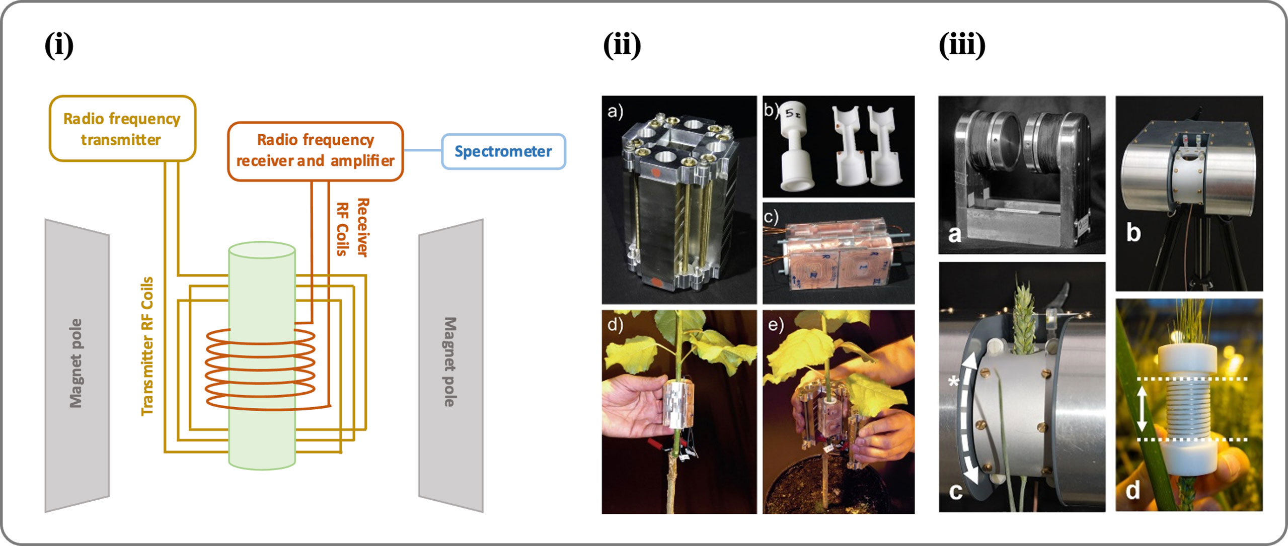 FIGURE 4 (I) An image showing the different parts of an NMR spectrometer. (II) Portable NMR CUFF magnet system (source: Windt et al., 2011). An NMR-CUFF prototype, consisting of a Halbach magnet, is shown in this image; (A) shown is the template for an RF-coil; (B) the hinged, plane-parallax gradient system assembly with an RF-coil inside; (C) mounting the RF coils; and (D) enclosing the plant stem with the NMR-CUFF. (III) Mobile NMR Mouse sensor made from a C-shaped magnet (source: Windt et al., 2021). An image of the magnet (A) shows a permanent C-shaped magnet, (B) an insulating housing or adjustable stand, (C) a probe housing that can be rotated around the magnet poles (*) to the sample angle, and (D) solenoidal RF coils with dotted lines indicating their sensitive volume (25 mm). For more details see Windt et al., 2021.
“